Gallbladder and Biliary Disease: Stones, Cholangitis, and ERCP Explained
Imagine a sudden, crushing pain in your upper right abdomen that hits like a freight train. It doesn’t just hurt; it leaves you breathless, sweating, and unable to find a comfortable position. For millions of people, this is the first time they realize their gallbladder is a small, pear-shaped organ under the liver that stores bile to help digest fats is failing them. This isn't just bad luck. Gallbladder and biliary diseases are among the most common reasons for hospital admissions in developed countries, affecting roughly 10-15% of adults. But what exactly is going on inside your body, and why do doctors talk so much about ERCP is Endoscopic Retrograde Cholangiopancreatography, a specialized procedure using endoscopy and X-rays to diagnose and treat bile duct issues?
Understanding these conditions requires looking beyond simple "stomach ache" diagnoses. We need to unpack how gallstones form, when they become dangerous enough to cause life-threatening infections like cholangitis, and why modern medicine relies heavily on minimally invasive techniques rather than open surgery. Let’s break down the anatomy, the risks, and the treatments without the confusing medical jargon.
How Gallstones Form and Why They Matter
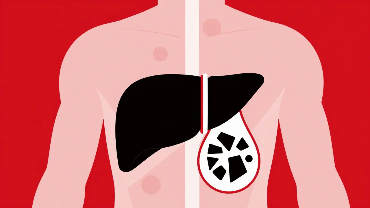
Your liver produces bile, a fluid essential for breaking down fats. The gallbladder stores this bile until you eat, then squeezes it into the intestines via the cystic duct and common bile duct. Problems start when the chemical balance of bile gets thrown off. If there is too much cholesterol, bilirubin, or calcium salts, these substances crystallize. Over time, they grow from microscopic grains into solid stones.
About 80% of these stones are made primarily of cholesterol. The other 20% are pigment stones, composed mainly of bilirubin, often seen in people with liver cirrhosis or blood disorders. Mixed stones contain both. The size varies wildly-from tiny specks to golf ball-sized masses. Here is the catch: having stones doesn't automatically mean you have a problem. Many people live with silent stones forever. However, if a stone blocks the cystic duct, it traps bile in the gallbladder, leading to inflammation known as acute cholecystitis. If it blocks the common bile duct further down, the consequences are far more severe, potentially causing jaundice, pancreatitis, or infection.
The Danger Zone: Understanding Cholangitis
When a gallstone lodges in the common bile duct or at the ampulla of Vater (where the bile duct meets the intestine), it creates a backup. Bile can't flow out, and bacteria from the intestine can travel up into the sterile bile ducts. This leads to acute cholangitis is a serious bacterial infection of the bile ducts caused by obstruction. This is not something you wait out at home. It is a medical emergency.
Doctors look for a specific set of symptoms called Charcot's triad to diagnose this condition:
- Right Upper Quadrant Pain: Present in about 70% of cases, this is intense pain under the right rib cage.
- Fever: Occurring in 85% of patients, indicating an active infection.
- Jaundice: Yellowing of the skin and eyes, seen in 60-70% of cases due to backed-up bile.
If the infection worsens, it can progress to Reynolds' pentad, which adds septic shock (dangerously low blood pressure) and altered mental status. Without prompt treatment, cholangitis can be fatal. The goal of immediate care is to relieve the blockage and control the infection, usually through antibiotics and drainage procedures.
Diagnosing Biliary Issues: From Ultrasound to MRCP
You don't need a guesswork diagnosis. Modern imaging is highly accurate. The first step is almost always a right upper quadrant ultrasound. It’s non-invasive, cheap, and effective. Ultrasound has a sensitivity of 84% and specificity of 96% for detecting gallstones in the gallbladder itself. However, ultrasounds struggle to see deep inside the bile ducts because gas in the intestines blocks the view.
If doctors suspect a stone is stuck in the bile duct-based on blood tests showing elevated liver enzymes or bilirubin-they move to Magnetic Resonance Cholangiopancreatography (MRCP is a specialized MRI technique that provides detailed images of the bile and pancreatic ducts without radiation). MRCP is the gold standard for diagnosis because it offers a clear map of the biliary tree with 95% sensitivity and 97% specificity for common bile duct stones. Crucially, it carries no risk of complications, unlike therapeutic procedures. Guidelines now strongly recommend using MRCP before attempting any invasive intervention to confirm the presence of stones.
ERCP: The Therapeutic Workhorse
This brings us to ERCP is Endoscopic Retrograde Cholangiopancreatography, a procedure combining endoscopy and fluoroscopy to diagnose and treat bile and pancreatic duct problems. While MRCP is for looking, ERCP is for acting. Developed in the late 1960s and refined significantly since the 1990s, ERCP allows gastroenterologists to reach the opening of the bile duct in the duodenum, inject dye to visualize the ducts under X-ray, and perform interventions.
The primary use of ERCP today is therapeutic. If MRCP confirms a stone in the common bile duct, the doctor performs an endoscopic sphincterotomy-a small cut in the muscle surrounding the duct opening-to allow the stone to pass or be extracted with a basket or balloon. Success rates exceed 90% in experienced hands. However, ERCP is not without risks. The most common complication is post-ERCP pancreatitis, occurring in 3-10% of cases. Other risks include bleeding, perforation of the intestine, and infection. Because of these risks, current guidelines dictate that ERCP should rarely be used for diagnosis alone. It is reserved for when treatment is needed.
Treatment Options: Surgery vs. Medication
Once the immediate crisis of a blocked duct is managed, the long-term solution usually involves removing the gallbladder. Laparoscopic cholecystectomy is a minimally invasive surgical procedure to remove the gallbladder using small incisions and a camera is the gold standard. It accounts for 90% of all gallbladder removals. Compared to open surgery, it offers massive advantages: hospital stays drop from nearly five days to just over one day, and recovery time shrinks from weeks to roughly 7-10 days. Post-operative pain is significantly reduced, and scarring is minimal.
Are there alternatives? Yes, but they are limited. Ursodeoxycholic acid (UDCA) is a medication that can dissolve small cholesterol stones. However, it takes 6-12 months to work, only succeeds in 30-40% of cases, and stones often return after stopping the drug. It is generally reserved for patients who cannot undergo surgery. Shock wave lithotripsy, which breaks stones with sound waves, has largely fallen out of favor due to high recurrence rates (50% within five years) and limited effectiveness for multiple stones.
| Modality | Primary Use | Sensitivity/Efficacy | Risk Level | Recovery Time |
|---|---|---|---|---|
| Ultrasound | Initial Diagnosis | 84% Sensitivity | None | N/A |
| MRCP | Detailed Imaging | 95% Sensitivity | None | N/A |
| ERCP | Therapeutic Intervention | >90% Success | Moderate (3-10% complications) | 1-2 Days |
| Laparoscopic Cholecystectomy | Gallbladder Removal | Gold Standard | Low-Moderate | 7-10 Days |
| UDCA Therapy | Stone Dissolution | 30-40% Success | Very Low | 6-12 Months |
Living With a Removed Gallbladder
After surgery, your body adapts. Without a storage tank, bile drips continuously from the liver into the intestine. Most people adjust quickly. About 75% of patients resume a normal diet within six weeks. Initially, a low-fat diet is recommended for 2-4 weeks to prevent digestive upset. Some patients experience persistent diarrhea or abdominal pain, known as post-cholecystectomy syndrome, affecting roughly 12% of individuals. This may require additional management or investigation to rule out retained stones or other causes.
The key takeaway is prevention where possible. Risk factors for gallstone formation include obesity (BMI >30 increases risk 2-3 fold), rapid weight loss, diabetes, and certain genetic predispositions. Women are affected 2.1 times more frequently than men. By maintaining a healthy weight and avoiding crash diets, you can significantly lower your risk of developing symptomatic stones.
Is ERCP painful?
During the procedure, you are under sedation or general anesthesia, so you feel no pain. Afterward, many patients report mild throat soreness from the scope and some abdominal bloating or discomfort. Most people return to light activities within 48 hours. Serious pain could indicate a complication like pancreatitis and should be reported to your doctor immediately.
Can I avoid surgery if I have gallstones?
If your gallstones are asymptomatic (silent), surgery is usually not recommended because the risk of complications from the operation outweighs the low annual risk of developing symptoms (1-2%). However, if you have symptomatic stones, recurrent attacks, or complications like pancreatitis, laparoscopic cholecystectomy is the standard and most effective treatment to prevent future emergencies.
What is the difference between MRCP and ERCP?
MRCP is purely diagnostic-it uses magnetic resonance imaging to create detailed pictures of your bile ducts without any invasion or risk. ERCP is both diagnostic and therapeutic, involving an endoscope and X-rays, allowing doctors to remove stones or place stents. Because ERCP carries a risk of complications, MRCP is typically done first to confirm the need for ERCP.
How long does it take to recover from gallbladder removal?
Most patients go home the same day or the next morning. You can usually return to desk work within a week. Strenuous activity should be avoided for about two weeks. Full internal healing takes longer, but most people feel back to normal within 7-10 days. Dietary adjustments may be needed for several weeks as your body adjusts to continuous bile flow.
What are the signs of cholangitis?
The classic signs are Charcot's triad: severe pain in the upper right abdomen, fever, and jaundice (yellowing of the skin or eyes). If you experience these symptoms together, seek emergency medical attention immediately. Untreated cholangitis can lead to sepsis, organ failure, and death.