Liver Function Tests Explained: Understanding ALT, AST, and Bilirubin Results

You walk into the clinic, get a blood draw, and weeks later, you hold a sheet of paper with columns of numbers. Some have arrows pointing up or down. You Google the results, find scary headlines about liver failure, and suddenly you think your favorite weekend beer caused permanent damage. Take a breath. Blood work looks complicated until you know the story behind the codes. Today, we are decoding Liver Function Tests (LFTs) so you understand exactly what is happening inside your body without needing a medical degree.
Most people call them liver tests, but doctors know they aren't always accurate indicators of how well your liver functions. Instead, they are often markers of damage. Think of it like a car warning light. If your engine temperature gauge spikes, the light tells you something is broken, but not necessarily which part. These tests measure enzymes leaking out of damaged cells or waste products building up because the filtering system is backed up. By early 2026, we still rely heavily on the standard panel that has been refined over decades, starting from the biochemical basics established by researchers in the 1970s.
The Core Enzymes: ALT and AST
Two numbers dominate most reports: ALT and AST. These are enzymes found inside liver cells. When those cells get hurt, they break open and spill these chemicals into your bloodstream. That is why your blood shows higher levels. It isn't that your liver makes too much enzyme; it's that the cell walls became too porous to keep them inside.
Alanine Aminotransferase (ALT) is an enzyme primarily located in the liver. Because it lives mostly there, high ALT usually points directly to liver injury. For example, if you take a heavy dose of acetaminophen by mistake, your ALT shoots up fast. Normal ranges vary slightly by lab, but generally, it sits between 7 and 55 units per liter (U/L). In men, it might sit a bit lower than women, though obesity often pushes these baselines higher.
Aspartate Aminotransferase (AST) is an enzyme found in the liver, heart, and muscles. This overlap matters. If you run a marathon three days before the test, your AST might look high due to muscle breakdown, tricking you into thinking your liver is sick. However, in liver contexts, AST reacts quickly. Its half-life is shorter than ALT's, meaning it spikes faster after acute injury but also drops faster once healing starts. Typical normal values range from 8 to 48 U/L.
The magic happens when you compare them. Doctors calculate a ratio known as the De Ritis ratio. If your AST is more than twice as high as your ALT, especially if both are less than 300, it strongly suggests alcohol-related liver strain. Conversely, if ALT is higher than AST, it often signals non-alcoholic issues like viral hepatitis or fatty liver disease. In modern terms, we refer to fatty liver as Metabolic dysfunction-Associated Steatotic Liver Disease (MASLD) following the 2023 guidelines. In these cases, ALT typically exceeds AST.
| Test Component | Typical Range | Primary Clue |
|---|---|---|
| ALT SGPT | 7 - 55 U/L | Liver cell damage |
| AST SGOT | 8 - 48 U/L | Liver, heart, or muscle damage |
| Bilirubin | 3 - 17 μmol/L | Bile flow blockage |
Bilirubin: The Yellow Warning Sign
If you've ever heard someone say a patient was "jaundiced," you saw bilirubin in action. Bilirubin is a yellow pigment formed when old red blood cells break down. Your liver is supposed to filter it and send it into your bile so it leaves your body through stool. But if your liver is sluggish or blocked, it stays in the blood, turning skin and eyes yellow.
Total Bilirubin measures everything, while Direct Bilirubin specifically tracks the processed form ready for excretion. High total bilirubin often means obstruction-like a gallstone blocking the duct-or severe liver stress where processing fails. Levels above 17 μmol/L usually warrant investigation. Interestingly, Gilbert Syndrome is a harmless genetic condition affecting up to 7% of people, causing occasional spikes in indirect bilirubin with no actual damage to the organ.
Checking the Factory Output: Albumin and Clotting
Enzymes tell us about injury, but do they tell us if the liver is actually working? That is where Albumin comes in. Think of albumin as a protein the factory makes constantly. It keeps fluid from leaking out of vessels into your tissues. If your albumin is low (below 3.5 g/dL), it suggests long-term synthetic failure. Because the protein has a 20-day lifespan, it reflects chronic issues rather than sudden problems. Even if your ALT is normal today, low albumin could mean hidden cirrhosis developed over years.
We also track clotting factors, specifically Prothrombin Time (PT). Vitamin K-dependent proteins help blood clots form. The liver makes them. If the PT stretches longer than usual, it signals the liver isn't manufacturing these protectors fast enough. This becomes critical in acute liver failure. Unlike albumin, PT changes quickly (within hours), so it is the best marker for immediate danger.
The Supporting Cast: ALP and GGT
Sometimes the problem isn't the cells dying, but the drainage pipes clogging. This creates a pattern called cholestasis. Two key players show up here. Alkaline Phosphatase (ALP) lives in the bile ducts. If this rises significantly (more than 3 times normal), suspect a blockage. Note that ALP also lives in bones. Growing teenagers or people with bone fractures can have high ALP with perfectly healthy livers.
To confirm if the ALP spike is liver-related, we look at Gamma-Glutamyl Transferase (GGT). GGT increases alongside ALP when the source is hepatic. If ALP is high but GGT is normal, look at the bones instead. GGT is also incredibly sensitive to alcohol intake. Chronic drinkers often show elevated GGT even when other liver enzymes seem quiet. It is often the first line of defense in screening for substance abuse patterns in blood work.
Reading the Patterns Like a Detective
Doctors rarely look at one number in isolation. They look for shapes in the data. StatPearls documents a standard diagnostic approach: compare transaminases (ALT/AST) against alkaline phosphatase (ALP).
If ALT is 10 times higher than ALP, you have a hepatocellular pattern. The liver tissue itself is inflamed. Common culprits include viral hepatitis, autoimmune attacks, or medication toxicity. Conversely, if ALP is 3 times higher than ALT, you have a cholestatic pattern. The tubes are blocked. You might suspect gallstones, strictures, or cancer pressing on the bile ducts.
Mixed patterns exist too. Drug-induced injury often raises all markers, making diagnosis tricky. A study from Hepatology involving over 12,000 patients showed that adding fibrosis scores like FIB-4 to these basic tests improved accuracy for finding advanced scarring from 68% to 89%. This means combining your blood numbers with age and platelet counts gives a clearer picture than staring at the raw LFTs alone.
Why "Normal" Isn't Always Safe
Hundreds of thousands of people live with advanced fibrosis yet maintain borderline normal LFTs. The liver has massive reserve capacity. It can lose 70% of its mass and still process toxins efficiently enough to hide damage on a standard panel. Conditions like Nonalcoholic Steatohepatitis (NASH) or now termed MASLD often present with just isolated GGT elevation or mildly high ALT. A 2022 study found nearly 37% of primary care physicians ordered unnecessary scans for ALT levels between 41-80 U/L, fearing cancer or hardening when lifestyle tweaks were the actual solution.
Also, remember context. Muscle injuries raise AST. Severe exercise skews results. Obesity correlates with naturally higher baseline ALT and AST ranges-roughly 10-15% higher in people with a BMI over 30 compared to leaner individuals. Your baseline is unique. One-off numbers matter less than trends over time.
Frequently Asked Questions
Can I fix abnormal liver enzymes by changing my diet?
Yes, in many metabolic cases like MASLD. Losing 10% of your body weight can significantly reduce fat accumulation in the liver, lowering ALT and AST levels naturally over months. Avoid sugar-sweetened drinks and prioritize fiber-rich foods.
What level of ALT indicates liver failure?
High ALT alone doesn't define failure. Failure is defined by low albumin and high prothrombin time indicating lost synthetic function. Extremely high ALT (>1000) usually suggests acute shock to the liver, such as from toxin exposure, but recovery is often possible.
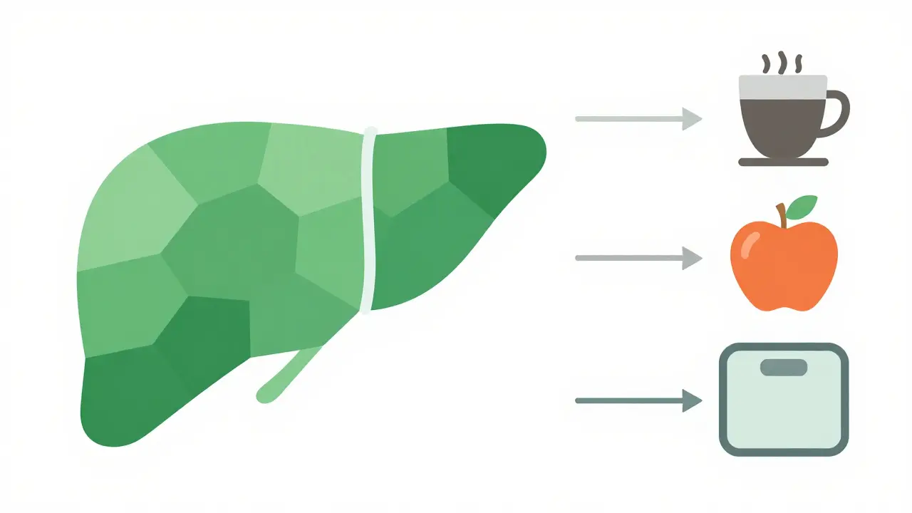
Does drinking coffee affect liver function tests?
Surprisingly, moderate coffee consumption has been linked to lower risks of liver fibrosis and cirrhosis. However, avoid energy drinks or excessive sugar mixes which can spike triglycerides and indirectly worsen liver fat storage.
Why did my doctor order an ultrasound after my blood test?
Blood tests reveal chemical imbalances, but imaging reveals structural issues. Ultrasound is excellent for spotting fatty deposits, gallstones, or masses that enzymes alone cannot locate precisely.
How fast do liver numbers return to normal?
AST clears faster (days) than ALT (weeks) due to half-life differences. Alcohol withdrawal might normalize GGT in 4-6 weeks. Viral hepatitis recovery varies, but acute spikes usually drop within months if inflammation stops.
Understanding these tests empowers you to discuss options confidently. Don't panic at a single arrow. Look at the pattern, the history, and the supporting evidence. Your liver is resilient, but monitoring it helps keep that resilience intact for the long haul.
Dan Stoof
March 30, 2026 AT 10:47Wow! The clarity here is absolutely fantastic!!! I finally feel like I am understanding what those arrows mean!!! The explanation about the car warning light is so creative and helpful!!! I never realized the difference between injury markers and function tests!!! It is truly enlightening!!!
Calvin H
March 31, 2026 AT 07:01Glad to know my nightly beverage isn't going to kill me apparently.
Adryan Brown
April 1, 2026 AT 05:50I have been reading through this extensively and the breakdown of enzymes is quite fascinating in its complexity. It is remarkable how the body manages these chemical balances without us noticing most of the time. The distinction between acute injury and chronic dysfunction is something many patients miss entirely. We often panic when we see a single red arrow on our lab results sheet. However, the context of the De Ritis ratio provides a much clearer diagnostic path forward. Understanding the half-life of AST versus ALT really changes how one interprets a sudden spike in numbers. The mention of muscle exertion affecting AST levels is a crucial piece of information often overlooked. It is easy to mistake a heavy gym session for a liver problem without knowing this nuance. Bilirubin levels can also be misleading if one does not account for genetic conditions like Gilbert Syndrome. The factory analogy regarding albumin production paints a vivid picture of synthetic capacity. We should remember that the liver has a massive reserve to handle stress before failure occurs. This resilience is both a blessing and a curse because damage hides until it is severe. The inclusion of clotting factors adds another layer of urgency for acute assessments. Chronic issues show up differently than sudden trauma events in the blood panel. Monitoring trends over time is far superior to worrying about a one-time snapshot. I hope more people read this and stop spiraling immediately after their next blood draw. Education is truly the best medicine when dealing with confusing medical data.
dPhanen DhrubRaaj
April 2, 2026 AT 23:24in india we see this often with jaundice cases but people panic too much sometimes the liver heals itself well if given rest and less alcohol
Cameron Redic
April 2, 2026 AT 23:30Actually the study cited from Hepatology regarding fibrosis scores needs more scrutiny. The sample size might be biased toward specific demographics. Relying solely on FIB-4 without imaging can lead to false positives in certain populations.
William Rhodes
April 3, 2026 AT 08:32You are missing the philosophical implication of how we view our own mortality through these numbers. It is not just about medical accuracy but about the anxiety of uncertainty. We crave absolute truth in biological systems that are inherently probabilistic. The desire to control health outcomes through numbers is a human flaw. True wisdom accepts the ambiguity that exists in every medical report.
Biraju Shah
April 4, 2026 AT 18:19The separation of cholestatic and hepatocellular patterns is vital for proper diagnosis. Patients must demand more than just a reference range comparison. Doctors need to explain the pattern, not just the individual value.
RONALD FOWLER
April 6, 2026 AT 09:54thank you for sharing this valuable information with the community here
Carolyn Kask
April 7, 2026 AT 10:49Our healthcare system finally producing content like this is a relief compared to other nations struggling with basic education. American standards are still leading the way in patient transparency.
Vikash Ranjan
April 9, 2026 AT 06:04I disagree with the notion that we are doing better elsewhere. Everyone deals with fatigue and liver stress equally. The barriers to entry for testing remain high for lower income groups globally.
Brian Yap
April 9, 2026 AT 12:49Nah mate, coffee is actually pretty protective for the liver if you drink it black. Just keep away from the sugary energy drinks that wreck your metabolism. Cheers to a healthy future!
Marwood Construction
April 11, 2026 AT 08:47This documentation aligns with occupational health safety protocols we implement for workers handling chemical exposures regularly. The distinction between enzyme leakage and synthetic function is critical for workplace fitness evaluations.
Kendell Callaway Mooney
April 11, 2026 AT 17:08Yes, simple terms help patients understand the basics quickly. Breaking down complex biochemistry into factory analogies works wonders for retention. It empowers individuals to advocate for themselves during appointments.
Katie Riston
April 13, 2026 AT 02:53There is a profound irony in seeking certainty from organs designed to process toxins constantly. The liver works silently while we wonder if it is failing us. Perhaps we should trust the body's innate resilience more than the arbitrary thresholds set by labs. Health is a journey rather than a static state of perfect numbers. We often forget that the body heals if we stop attacking it with sugar and alcohol.
Charles Rogers
April 13, 2026 AT 17:53Most people ignore the ratios until it is too late to prevent fibrosis progression. They focus on ALT but miss the Albumin dropping slowly. Early intervention requires monitoring the factory output markers proactively rather than waiting for cell death signals. Prevention beats cure in every metabolic scenario presented here.